Liver: organ not to be overloaded

NON-ALCOHOLIC HEPATIC STEATOSIS
Non-alcoholic fatty liver disease (NAFLD) is the most common form of chronic liver disease worldwide (about 25% of the general population), and among patients, a not insignificant percentage (3-5%) develops non-negligible steatohepatitis. alcoholic (NASH), characterized by damage to hepatocytes, inflammation and fibrosis, which increase the risk of developing cirrhosis and hepatocellular carcinoma.
At the base of the pathogenesis of NAFLD there is an accumulation of fats, in particular of triglycerides (TG) in the liver. It is therefore not surprising that most people with NAFLD have conditions such as visceral obesity (fat accumulation within the waist), hypertension, hyperlipidemia, and diabetes.
However, the mechanisms by which a minority of patients develop a more severe phenotype than others are still not fully understood. In a recent review carried out by the University of Perugia and the Polytechnic of Ancona, together with the Scientific Division of Lipinutragen, it is clear that, in addition to the accumulation of TG, the main cause of the progression of the steatotic form in steatohepatitis was determined in the LIPOTOXICITY, or in the accumulation of specific classes of lipids (free fatty acids, ceramides, cholesterol, bile acids) which act as harmful agents [1]. These species, defined as “lipotoxic”, influence cell behavior through multiple mechanisms that induce the activation of cell death and oxidative stress receptors.
ROLE OF FREE FATTY ACIDS (FFA) IN LIPOTOXICITY
The most studied mechanisms underlying the progression from NAFLD to NASH are those that lead to the release and transformation of lipid species defined as “neutral” (i.e. TG) into Free Fatty Acid (FFA).
In particular, it appears that more than the quantity of FFA that accumulate in the liver, it is their quality that influences the lipotoxic events.
Among these, saturated fatty acids (SFA), such as palmitic acid and stearic acid, have shown greater toxicity than monounsaturated fatty acids (MUFA), such as oleic acid, which instead show a protective role for the cell [2]. Along with MUFAs, polyunsaturated fatty acids (PUFAs) of the omega-3 series also have a proven protective activity for the liver. As evidenced by recent clinical studies [3-5], omega-3 PUFAs, in addition to reducing TG levels in plasma and liver, reduce transaminase (ALT) values and in general lead to an improvement in ultrasound parameters in liver in both adult and pediatric NASH patients.
Conversely, an accumulation of PUFA of the omega-6 series, and in particular of arachidonic acid (AA), favors the formation of inflammatory molecules, charging the process of lipotoxicity.
EVIDENCE OBTAINED WITH THE LIPIDOMIC ANALYSIS OF MATURE ERYTHROCYTES
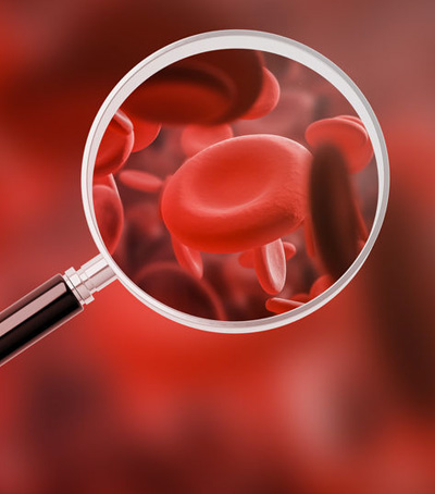 The growing understanding of these lipid anomalies through lipidomic technologies, such as membrane lipidomic analysis of mature red blood cells (GRM), enrich the understanding of the phenomenon by directly evaluating the presence of lipotoxicity as a key pathogen at the cellular level.
An important point in favor of the use of erythrocytes, as a reporter for liver disease, is the similarity of their composition in terms of membrane fatty acids with that of hepatocytes:
• SFA: 43% in the GRM versus 42% in the liver
• MUFA: 23.0% in the GRM versus 23.8% in the liver
• Omega-6 PUFA: 27.6 in the GRM versus 27.4% in the liver
• Omega-3 PUFA: 5.7% in the GRM versus 4.6% in the liver [6].
The average life span of cells, 120 days for erythrocytes and 180 days for hepatocytes, is also a clear indication of similar metabolism among the fatty acids that make up the phospholipids of their membranes.
SCIENTIFIC EVIDENCE AND ROLE OF NUTRITION
Recent studies report the possibility of treating NAFLD and NASH subjects by exploiting the benefits of a dietary supplement, in particular with vitamin E and PUFA of the omega-3 series [7-9]. Vitamin E, in fact, represents the most important molecule “for blocking” the oxidative cascade caused by free radicals. In fact, it is now recommended in clinical guidelines of the EU, US and Asia as a therapy for NASH in non-diabetic patients [8-9]. While, as regards the omega-3 PUFAs, DHA represents a promising molecule to prevent the progression of the disease to NASH [7] thanks to its anti-inflammatory activity also acting at the level of the membranes, improving their plasticity and fluidity.
From these premises, clinical studies have been carried out that have also explored the potential of multivitamin formulations containing DHA, as the main component, in the therapy of pediatric NASH. The results show a marked improvement in the metabolic, ultrasound and histological parameters of the liver. The results of this and other studies confirm the importance of PUFA as biomarkers for a non-invasive diagnosis of NASH, describing for the first time its importance for verifying the effectiveness of what we can define as a “DHA therapy”. In light of the above, it would be even more important to customize the lipid therapy and also to follow it as efficacy, both clinical and molecular, over time by performing the membrane lipidomic analysis of the GRM. The knowledge is there, the diagnostic tool too, so it is only time to adapt the protocols, taking into account that the analysis is now performed by the Lipidomics Lipinutragen Laboratory with an ISO 17025 accredited method, therefore with the appropriate specifications for the certainty of the data obtained.
Bibliography:
• G. Svegliati-Baroni, I. Pierantonelli, P. Torquato, R. Marinelli, C. Ferreri,
C. Chryssostomos, D. Bartolini., F. Galli. Lipidomic biomarkers and mechanism of lipotoxicity in non-alcoholic fatty liver disease, Free Radical Biology and Medicine 144 (2019) 293–299.
• Y. Akazawa, S. Cazanave, J.L. Mott, N. Elmi, S.F. Bronk, S. Kohno, M.R. Charlton, G.J. Gores.
Palmitoleate attenuates palmitate-induced Bim and PUMA up-regulation and hepatocyte lipoapoptosis. J. Hepatol., 52 (4) (2010), pp. 586-593.
• C. Della Corte, G. Carpino, R. De Vito, C. De Stefanis, A. Alisi, S. Cianfarani, D. Overi, A. Mosca, L. Stronati, S. Cucchiara, M. Raponi, E. Gaudio, C.D. Byrne, V. Nobili. Docosahexanoic acid plus vitamin D treatment improves features of NAFLD in children with serum vitamin D deficiency: results from a single centre trial. PLoS One, 11 (12) (2016), Article e0168216.
• E. Zohrer, A. Alisi, J. Jahnel, A. Mosca, C. Della Corte, A. Crudele, G. Fauler, V. Nobili.
Efficacy of docosahexaenoic acid-choline-vitamin E in paediatric NASH: a randomized controlled clinical trial. Appl. Physiol. Nutr. Metabol., 42 (9) (2017), pp. 948-954.
• P. Torquato, D. Giusepponi, A. Alisi, R. Galarini, D. Bartolini, M. Piroddi, L. Goracci, A. Di Veroli, G. Cruciani, A. Crudele, V. Nobili, F. Galli. Nutritional and lipidomics biomarkers of docosahexaenoic acid-based multivitamin therapy in pediatric NASH. Sci. Rep., 9 (1) (2019), p. 2045.
• L. Lauritzen, H.S. Hansen, M.H. Jorgensen, K.F. Michaelsen. The essentiality of long chain n-3 fatty acids in relation to development and function of the brain and retina. Prog. Lipid Res., 40 (1–2) (2001), pp. 1-94
• V. Nobili, A. Alisi, G. Musso, E. Scorletti, P.C. Calder, C.D. Byrne. Omega-3 fatty acids: mechanisms of benefit and therapeutic effects in pediatric and adult NAFLD. Crit. Rev. Clin. Lab. Sci., 53 (2) (2016), pp. 106-120.
• F. Galli, A. Azzi, M. Birringer, J.M. Cook-Mills, M. Eggersdorfer, J. Frank, G. Cruciani, S. Lorkowski, N.K. Ozer. Vitamin E: emerging aspects and new directions.
• V.W. Wong, S. Chitturi, G.L. Wong, J. Yu, H.L. Chan, G.C. Farrell. Pathogenesis and novel treatment options for non-alcoholic steatohepatitis. Lancet Gastroenterol. Hepatol., 1 (1) (2016), pp. 56-67.
Article by the Editorial team of Lipinutragen
The information given must in no way replace the direct relationship between health professional and patient.
Photo: 123RF Archivio Fotografico: 112228944 ©lightfieldstudios / 123rf.com | 106657631 ©ismagilov / 123rf.com
- On 15 January 2021
